- ALL COMPUTER, ELECTRONICS AND MECHANICAL COURSES AVAILABLE…. PROJECT GUIDANCE SINCE 2004. FOR FURTHER DETAILS CALL 9443117328
Projects > COMPUTER > 2019 > NON IEEE > APPLICATION
Automatic based kidney segmentation is proposed in this research. In biometric or medical image processing the segmentation of kidney probably take several hours. This should be overcome by applying the multi thread technology. The best part is to divide the kidney segmentation into four components. The segmented components are renal cortex, renal column, renal medulla and renal pelvis. It is further divided into two components they are localization of renal cortex, and segmentation of kidney components. The localization of renal cortex is done with the help of Generalized Hough Transform (GHT) and Active Appearance Model (AAM). The segmentation of kidney component is done by the help of modified Random Forest (RF). The randomized Forest component segment the kidney into four components based on the result of localization phase. Among all these technology multi-threading concept is included to speed up the process. Thus the segmentation result is provided more accurately than the previous concept of applied methodology.
In previous mechanism semi-automatic mechanism is implemented to segment the kidney. The method they introduced is named as graph cut method which is time consuming. Then lately they introduce time intensity curves but to read the spatial information using this is inappropriate. And the overall method they used is not effective. Kidney and renal cortex segmentation in CT, MRI and Ultrasound images include both semi-automatic and fully automatic methods. Freiman, Ali and Chen applied graph cut based method. Xie segmented kidney from Ultrasound images based on shape and texture priors. For kidney segmentation in dynamic MR images, not only the spatial information but also the timing activities, also known as time intensity curves, were used for kidney segmentation. Cuingnet used Random Forests to detect and segment kidneys in 3D CT images.
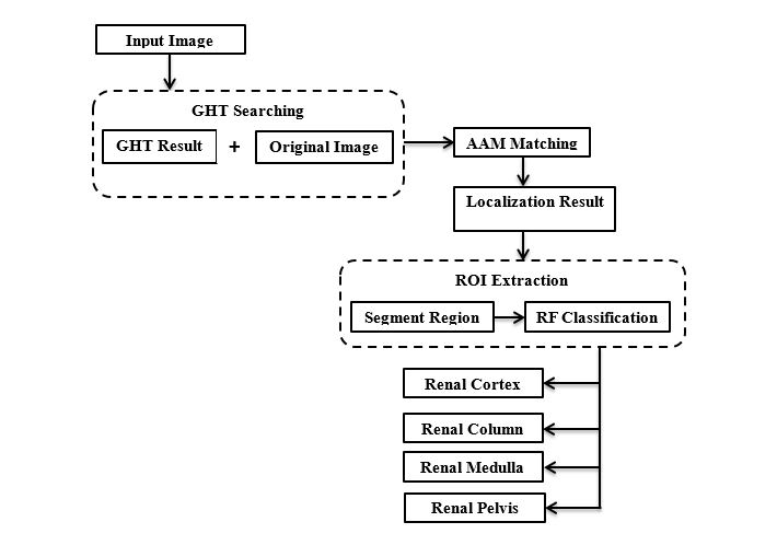
Localization and segmentation of kidney is proposed in this research work. In the localization of renal cortex phase, a method which combines Generalized Hough Transform (GHT) and Active Appearance Models (AAM) is applied to localize the kidney and estimate the thickness of renal cortex. In the segmentation phase, a modified Random Forests (RF) method is proposed to segment the kidney into multiple components based on the result from localization phase. During the segmentation process, renal cortex thickness table and thick constraint model are built, which are used to help the segmentation of cortex and column. Then an accumulator matrix indicating the possible position of the object in constructed according to the R-table. The AAM method matches a new data to the appearance model through minimizing the root mean square (RMS) intensity between the new data and appearance model instance by modifying the affine transformation, global intensity parameters, and appearance coefficients. Before segmentation, the volume of interest for the kidney is extracted based on the localization result. In the RF classification, the renal cortex and renal column are considered as one category because their intensity and texture are very similar. After this process, renal cortex, renal column, renal medulla and renal pelvis as well as background are segmented. Morphological methods are also applied. Opening operation is applied to remove isolated points, followed by closing operation to fill small holes.
ARCHITECTURE DIAGRAM