- ALL COMPUTER, ELECTRONICS AND MECHANICAL COURSES AVAILABLE…. PROJECT GUIDANCE SINCE 2004. FOR FURTHER DETAILS CALL 9443117328
Projects > ELECTRONICS > 2018 > IEEE > DIGITAL IMAGE PROCESSING
Recent advances in optical coherence tomography (OCT) lead to the development of OCT angiography to provide additional helpful information for diagnosis of diseases like basal cell carcinoma. In this paper, we investigate how to extract blood vessels of human skin from full-field OCT (FF-OCT) data using the robust principal component analysis (RPCA) technique. Specifically, we propose a short-time RPCA method that divides the full-field OCT data into segments and decomposes each segment into a low-rank structure representing the relatively static tissues of human skin and a sparse matrix representing the blood vessels. The method mitigates the problem associated with the slow varying background and is free of the detection error that RPCA may have when dealing with FF-OCT data. Both short-time RPCA and RPCA methods can extract blood vessels from FF-OCT data with heavy speckle noise, but the former takes only half the computation time of the latter. We evaluate the performance of the proposed method by comparing the extracted blood vessels with the ground truth vessels labeled by a dermatologist and show that the proposed method works equally well for FF-OCT volumes of different quality. The average F-measure improvements over the correlation-mapping OCT method, the modified amplitude-decorrelation OCT angiography method, and the RPCA method, respectively, are 0.1835, 0.1032, and 0.0458.
Robust principle component analysis (RPCA)
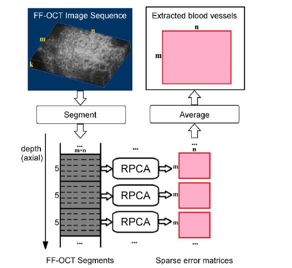
In light of its strength for background subtraction, the RPCA technique is employed in the proposed method to extract blood vessels from FF-OCT data. The red blood cells, which are sparsely distributed in the en face images, move in the blood vessels while other parts of the skin tissue remain nearly static across consecutive en face images. Therefore, the red blood cells can be considered as foreground and the other parts of the skin tissue as background. In a typical RPCA formulation, the background is modeled as a low-rank matrix and the foreground as a sparse matrix. Using this formulation, we vectorize each face image and represent the entire FF-OCT volume as a 2-D matrix. Then, we decompose the matrix into a low-rank matrix representing the relatively static part of the skin tissue and a sparse matrix representing the blood vessels.
BLOCK DIAGRAM