- ALL COMPUTER, ELECTRONICS AND MECHANICAL COURSES AVAILABLE…. PROJECT GUIDANCE SINCE 2004. FOR FURTHER DETAILS CALL 9443117328
Projects > ELECTRONICS > 2018 > IEEE > MEDICAL IMAGE PROCESSING
Robust organ segmentation is a prerequisite for computer-aided diagnosis (CAD), quantitative imaging analysis, pathology detection and surgical assistance. For organs with high anatomical variability (e.g., the pancreas), previous segmentation approaches report low accuracies, compared to well studied organs, such as the liver or heart. We present an automated bottom up approach for pancreas segmentation in abdominal computed tomography (CT) scans. The method generates a hierarchical cascade of information propagation by classifying image patches at different resolutions and cascading (segments) superpixels. The system contains four steps: 1) decomposition of CT slice images into a set of disjoint boundary-preserving superpixels; 2) computation of pancreas class probability maps via dense patch labeling; 3) superpixel classification by pooling both intensity and probability features to form empirical statistics in cascaded random forest frameworks; and 4) simple connectivity based post-processing. Dense image patch labeling is conducted using two methods: efficient random forest classification on image histogram, location and texture features; and more expensive (but more accurate) deep convolutional neural network classification, on larger image windows (i.e., with more spatial contexts). Over segmented 2D CT slices by the Simple Linear Iterative Clustering (SLIC) approach are adopted through model/parameter calibration and labeled at the superpixel level for positive (pancreas) or negative (non-pancreas or background) classes. The proposed method is evaluated on a dataset of 80 manually segmented CT volumes, using six-fold cross-validation. Its performance equals or surpasses other state-of-the-art methods (evaluated by “leave-one-patient-outâ€), with a Dice coefficient of 70:7% and Jaccard Index of 57:9%. In addition, the computational efficiency has improved significantly, requiring a mere 6 _ 8 minutes per testing case, versus _ 10 hours for other methods. The segmentation framework using deep patch labelling confidences is also more numerically stable, as reflected in the smaller performance metric standard deviations. Finally, we implement a multi-atlas label fusion (MALF) approach for pancreas segmentation using the same dataset. Under six-fold cross-validation, our bottom-up segmentation method significantly outperforms its MALF counterpart: 70:7 _ 13:0% versus 52:51 _ 20:84% in Dice coefficients.
Three-phase contrast enhanced CT data, multi-organ segmentation by combining inter-organ spatial interrelations with probabilistic atlases.
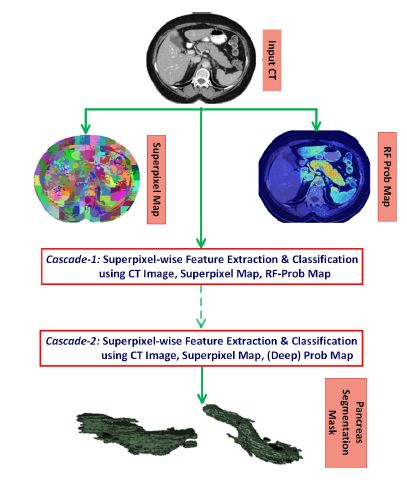
This paper proposes a new, bottom-up method using image and (deep) patch-level labeling confidences for pancreas segmentation. The method is applied to 80 single phase CT patient volumes. The method is designed to improve the segmentation accuracy of highly deformable organs, like the pancreas, by leveraging the middle-level representation of image segments. First, all 2D slices of an input patient abdominal CT scan are over-segmented into superpixels. Second, the superpixels are classified into two semantic classes (pancreas and nonpancreas) using multi-stage feature extraction and a random forest (RF) classification process on the image and (deep) patch-level confidence maps, pooled at the superpixel level. Two cascaded random forest superpixel classification frameworks are presented and compared.
BLOCK DIAGRAM