- ALL COMPUTER, ELECTRONICS AND MECHANICAL COURSES AVAILABLE…. PROJECT GUIDANCE SINCE 2004. FOR FURTHER DETAILS CALL 9443117328
Projects > ELECTRONICS > 2018 > IEEE > MEDICAL IMAGE PROCESSING
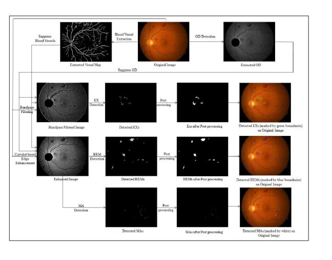
Diabetic Retinopathy (DR) is characterized by the progressive deterioration of retina with the appearance of different types of lesions that include microaneurysms, hemorrhages, exudates etc. Detection of these lesions plays significant role for early diagnosis of DR. To this aim, this paper proposes a novel and automated lesion detection scheme which consists of the four main steps: vessel extraction and optic disc removal, pre-processing, candidate lesion detection and post-processing. The optic disc and the blood vessels are suppressed first to facilitate further processing. Curvelet based edge enhancement is done to separate out the dark lesions from the poorly illuminated retinal background while the contrast between the bright lesions and the background is enhanced through an optimally designed wideband bandpass filter. The mutual information of the maximum matched filter response and the maximum Laplacian of Gaussian response are then jointly maximized. Differential Evolution algorithm is used to determine the optimal values for the parameters of the fuzzy functions that determine the thresholds of segmenting the candidate regions. Morphology based post-processing is finally applied to exclude the falsely detected candidate pixels.
Support vector machine (SVM) and matched filter (MF).
This paper proposes a novel and automated lesion detection scheme which consists of the four main steps: vessel extraction and optic disc removal, pre-processing, candidate lesion detection and post-processing. The optic disc and the blood vessels are suppressed first to facilitate further processing. Curvelet based edge enhancement is done to separate out the dark lesions from the poorly illuminated retinal background while the contrast between the bright lesions and the background is enhanced through an optimally designed wideband bandpass filter. The mutual information of the maximum matched filter response and the maximum Laplacian of Gaussian response are then jointly maximized. Differential Evolution algorithm is used to determine the optimal values for the parameters of the fuzzy functions that determine the thresholds of segmenting the candidate regions. Morphology based post-processing is finally applied to exclude the falsely detected candidate pixels.
BLOCK DIAGRAM