- ALL COMPUTER, ELECTRONICS AND MECHANICAL COURSES AVAILABLE…. PROJECT GUIDANCE SINCE 2004. FOR FURTHER DETAILS CALL 9443117328
Projects > ELECTRONICS > 2018 > IEEE > MEDICAL IMAGE PROCESSING
Segmentation of brain tumors from magnetic resonance imaging (MRI) datasets is of great importance for improved diagnosis, growth rate prediction and treatment planning. However, automating this process is challenging due to the presence of severe partial volume effect and considerable variability in tumor structures, as well as imaging conditions, especially for the gliomas. In this work, we introduce a new methodology that combines random forests and active contour model for the automated segmentation of the gliomas from multimodal volumetric MR images. Specifically, we employ a feature representations learning strategy to effectively explore both local and contextual information from multimodal images for tissue segmentation by using modality specific random forests as the feature learning kernels. Different levels of the structural information is subsequently integrated into concatenated and connected random forests (ccRFs) for gliomas structure inferring. Finally, a novel multiscale patch driven active contour (mpAC) model is exploited to refine the inferred structure by taking advantage of sparse representation techniques. Results reported on public benchmarks reveal that our architecture achieves competitive accuracy compared to the state-of-the-art brain tumor segmentation methods while being computationally efficient.
Random forest (RF) and Active contour models (ACMs)
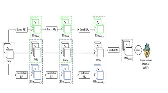
The proposed approach is able to integrate multiscale task-adapted information from multimodal MR volumes together for a fully automated, accurate and robust tissue segmentation. Specifically, we employ RFs as feature learning kernels to learn multiscale feature representations directly from multimodal MR volumes. The generated tissue feature maps are then fed into the subsequent concatenated classification forests to iteratively learn sequential tissue classifiers. The output tissue probability maps of a concatenated layer are further used as input augmented features to the subsequent concatenated layer. By connecting the estimations from multiscale concatenated classifiers and testing them on the trained global RFs, we can infer the brain tumor structure for a given testing subject. The inferred brain tumor structure is further incorporated into a multiscale patch driven active contour (mpAC) model as an initial contour as well as a spatial prior for the final segmentation. Compared to the previous methods using RF as the classifier for tissue segmentation, the proposed method allows the RF to do representation learning within a concatenated and connected architecture. Furthermore, by reformulating the voxel-wise classification of RFs as a contour evolution, our method achieves accurate and smooth segmentation of brain tumors. In addition, in contrast to the previous ACM methods, the mpAC refinement stage of our method is an automated one and exhibits more robustness to low tissue contrast. Validations on public available datasets have demonstrated significant advantages of the proposed method.
BLOCK DIAGRAM