- ALL COMPUTER, ELECTRONICS AND MECHANICAL COURSES AVAILABLE…. PROJECT GUIDANCE SINCE 2004. FOR FURTHER DETAILS CALL 9443117328
Projects > COMPUTER > 2019 > NON IEEE > APPLICATION
Liver segmentation on CT and MRI using laplacian mesh optimization is used to describe a semi-automated segmentation method for the liver and evaluate its performance on CT and MR images. The process starts by initializing an approximate 3D model from a few users are generated that contours to globally outline the liver shape. Then the model is then automatically deformed by a Laplacian mesh optimization scheme until it precisely delineates the patient’s liver. To improve the segmentation until satisfaction a correction tool was implemented to allow the user. The proposed method was tested against 30 CT-scans from the SLIVER07 challenge repository and 20 MR studies from the Montreal University Hospital Centre (CHUM), covering a wide spectrum of liver morphologies and pathologies. The average volumetric overlap error was 5.1% for CT and 7.6% for MRI and the average segmentation time was 6 minutes. The obtained results show that the proposed method is efficient, reliable and could effectively be used routinely in the clinical setting. The Significance of the proposed approach can alleviate the cumbersome and tedious process of slice-wise segmentation required for precise hepatic volumetry, virtual surgery and treatment planning.
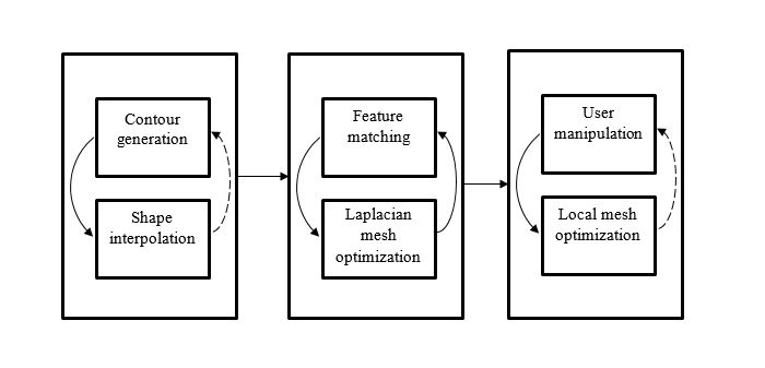
In the existing system, semi-automated segmentation method was used. First, an initial shape of the liver is generated by variational interpolation from a few user-generated contours. A template-matching method then dentifies target points corresponding to the liver boundary. Using a Laplacian mesh optimization framework, the geometric model is iteratively deformed until it converges to the liver boundary. Depending on the chosen treatment, careful surgical planning must be carried out. This involves assessment of liver volume prior to surgery to ensure sufficient residual liver function in major resections and in living-related liver transplantation. Traditionally, hepatic volumetry is performed by a radiology which manually delineates the liver on every slices of a CT-Scan
Automated liver segmentation is a challenging task in the field of medical image processing. In the proposed system, the Laplacian mesh optimization method was used. Initially the correction tool was developed. The proposed method consists of 3 main phases. First, an initial shape is interpolated from a few user-generated contours. This shape is then iteratively optimized to converge toward the liver boundary. Finally, the 3D surface mesh can be interactively manipulated to achieve the desired precision. A correction tool was implemented to improve the segmentation until satisfaction. The proposed approach can alleviate the cumbersome and tedious process of slice-wise segmentation required for precise hepatic volumetry, virtual surgery and treatment planning.
ARCHITECTURE DIAGRAM