- ALL COMPUTER, ELECTRONICS AND MECHANICAL COURSES AVAILABLE…. PROJECT GUIDANCE SINCE 2004. FOR FURTHER DETAILS CALL 9443117328
Projects > COMPUTER > 2019 > NON IEEE > APPLICATION
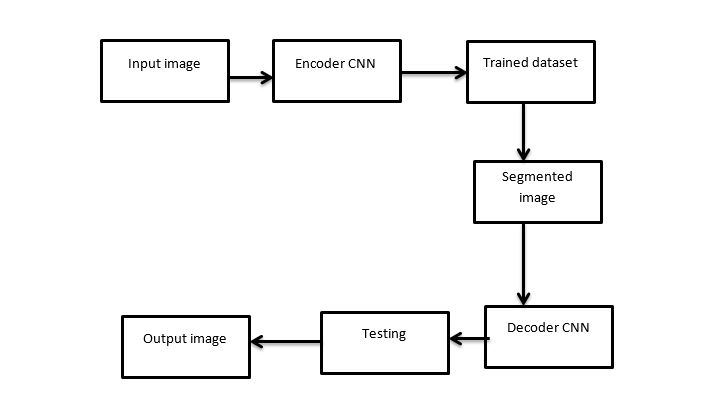
Tuberculosis (TB) is an infectious disease caused by the bacillus Mycobacterium tuberculosis. TB is a leading cause of death by infectious disease worldwide, alongside human immunodeficiency virus–acquired immune deficiency syndrome. The classification of tuberculosis were detected by using Deep Learning methods and Encoder-Decoder Convolutional Neural Networks (EDCNN). It performs the context of large-scale screening of population for lung and heart diseases as well as development of computational services for international portal on lung tuberculosis. It mainly performs two activities. The first activity aims to develop large-scale, real-world and well-annotated X-ray image database dedicated for automated TB screening. The second research activity focus on developing effective and efficient computational models to classify the image into different category of TB manifestations. The ED-CNN networks may be considered as a promising tool for automatic lung segmentation in large-scale projects.
Tuberculosis is a major health threat in many regions of the world. Opportunistic infections in immune compromised HIV/AIDS patients and multi-drug-resistant bacterial strains have exacerbated the problem, while diagnosing tuberculosis still remains a challenge. When left undiagnosed and thus untreated, mortality rates of patients with tuberculosis are high. Standard diagnostics still rely on methods developed in the last century. They are slow and often unreliable. In an effort to reduce the burden of the disease, it presents our automated approach for detecting tuberculosis in conventional postero anterior chest radiographs. It first extract the lung region using a graph cut segmentation method. For this lung region, we compute a set of texture and shape features, which enable the x-rays to be classified as normal or abnormal using a binary classifier. The system measure the performance of our system on two datasets: a set collected by the tuberculosis control program of our local county’s health department in the United States, and a set collected by Shenzhen Hospital, China. The computer-aided diagnostic system for TB screening, which is ready for field deployment, achieves a performance that approaches the performance of human experts. It achieve an area under the ROC curve (AUC) of 87% (78.3% accuracy) for the first set, and an AUC of 90% (84% accuracy) for the second set. For the first set, we compare our system performance with the performance of radiologists. When trying not to miss any positive cases, radiologists achieve an accuracy of about 82% on this set, and their false positive rate is about half of our system’s rate.
The chest radiographic images were resized to a 256 * 256 matrix and converted into Portable Network Graphics format. The images were loaded onto a computer with a Linux operating system dependencies for graphics processing unit acceleration. Two different deep convolutional neural network architectures were evaluated including pretrained and untrained models. Pretrained networks were already trained on 1.2 million everyday color images from ImageNet that consisted of 1000 categories before learning from the chest radiographs. Untrained networks were not trained before they were used referred to as untrained. Pretrained networks were obtained as, an open-access repository of pretrained models for use with Caffe. The following solver parameters were used for training: 120 epochs; base learning rate for untrained models and for pretrained models, 0.01 and 0.001, respectively; stochastic gradient descent; step-down, 33%; and g, 0.1. All images were augmented by using random cropping of 227 3 227 pixels, mean subtraction, and mirror images, which were prebuilt options. Further augmentation was performed in training some of the DCNNs, including rotations of 90°, 180°, and 270°, and Contrast Limited Adaptive Histogram Equalization processing by using Image. The DCNNs that used this additional augmentation are labeled when pretrained on ImageNet, and when untrained.
ARCHITECTURE DIAGRAM