- ALL COMPUTER, ELECTRONICS AND MECHANICAL COURSES AVAILABLE…. PROJECT GUIDANCE SINCE 2004. FOR FURTHER DETAILS CALL 9443117328
Projects > COMPUTER > 2019 > NON IEEE > APPLICATION
Automatic skin lesion segmentation is a challenging task due to the low contrast between lesion and the surrounding skin, the irregular and fuzzy lesion borders, the existence of various artifacts, and various imaging acquisition conditions. In this project, a fully automatic method for skin lesion segmentation by leveraging a 19-layer deep convolutional neural networks (CNNs) is trained end-to-end and does not rely on prior knowledge of the data. A set of strategies was proposed to ensure effective and efficient learning with limited training data. Furthermore, a novel loss function was designed based on Jaccard distance to eliminate the need of sample re-weighting, a typical procedure when using cross entropy as the loss function for image segmentation due to the strong imbalance between the number of foreground and background pixels.
A new technique for melanocytic lesion segmentation, Mimicking Expert Dermatologists Segmentations(MEDS), and extensive tests of its accuracy, speed, and robustness. MEDS combines a thresholding scheme reproducing the cognitive process of dermatologists with a number of optimizations that may be of independent interest. MEDS is simple, with a single parameter tuning its “tightnessâ€. It is extremely fast, segmenting medium resolution images in a fraction of a second even with the modest computational resources of a cell phone, an improvement of an order of magnitude or more over state-of-the-art techniques. And it is extremely accurate: very experienced dermatologists disagree with its segmentations less than they disagree with the segmentations of state-of-the-art techniques, and in fact less than they disagree with the segmentations of dermatologists of moderate experience. The first is optional and simply preprocesses the image to rebalance its colors andor to automatically remove any hair. The second stage reduces the dimensionality of the color space to 1 through Principal Component Analysis (PCA) of the color histogram. The third stage applies a blur filter to the resulting image to reduce noise. The fourth stage separates pixels into two clusters through a novel thresholding algorithm that is the heart of our technique and mimics the cognitive process of dermatologists, this effectively partitions the original image into regions corresponding to lesion and non lessional skin. The fifth stage morphologically post processes the image to remove spurious “patches†and to identify lesion areas of clinical interest it does so through a novel border detection scheme that appears at least 30% faster than the fastest existing schemes, and that may thus be of independent interest. Threshold based method rely on the histogram distribution of image color, which may undesirably altered in cases when significant amount of hair and bubbles are present.
Automatic skin lesion segmentation in dermoscopic images is a challenging task due to the low contrast between lesion and the surrounding skin, the irregular and fuzzy lesion borders, the existence of various artifacts, and various imaging acquisition conditions. In a fully automated method for skin lesion segmentation by leveraging the discriminative power of a 19-layer deep FCN. To the best of our knowledge, then it is among the first few attempts to use deep neural networks to tackle this challenging problem. We investigate a set of training strategies to ensure effective and efficient learning with limited training data. Secondly, we design an appropriate loss function that naturally handles the lesion-background imbalance of pixel wise classification for medical image segmentation. Our results show that this loss function can further improve the segmentation performance. At last, we extensively which evaluate the effectiveness, efficiency and the generalization capability of the proposed model using two large databases. Our model can be easily generalized to other challenging medical image segmentation problems.
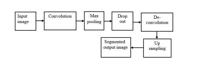
ARCHITECTURE DIAGRAM