- ALL COMPUTER, ELECTRONICS AND MECHANICAL COURSES AVAILABLE…. PROJECT GUIDANCE SINCE 2004. FOR FURTHER DETAILS CALL 9443117328
Projects > ELECTRONICS > 2019 > IEEE > DIGITAL IMAGE PROCESSING
This paper proposes a computer assisted diagnostic (CAD) system for the detection of melanoma in dermoscopy images. Clinical findings have concluded that in case of melanoma, the lesion borders exhibit differential structures such as pigment networks and streaks as opposed to normal skin spots, which have smoother borders. We aim to validate these findings by performing segmentation of the skin lesions followed by an extraction of the peripheral region of the lesion that is subjected to feature extraction and classification for detecting melanoma. For segmentation, we propose a novel active contours based method that takes an initial lesion contour followed by the usage of Kullback-Leibler divergence between the lesion and skin to fit a curve to the lesion boundaries. After segmentation of the lesion, its periphery is extracted to detect melanoma using image features that are based on local binary patterns. For validation of our algorithms, we have used the publicly available PH2 and ISIC dermoscopy datasets. An extensive experimental analysis reveals two important findings: 1. The proposed segmentation method mimics the ground truth data, and 2. The most significant melanoma characteristics in the lesion actually lie on the lesion periphery.
Gradient filter, Gabor filter and edge based filtering method.
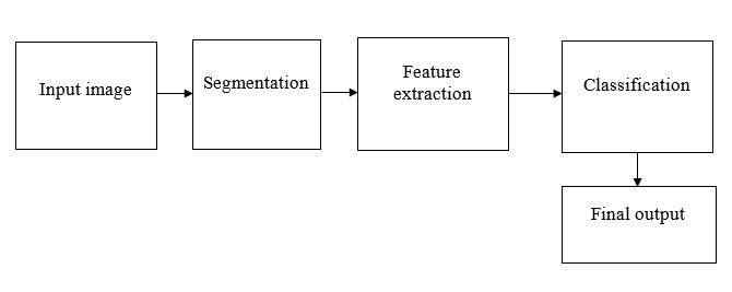
The main objective of this paper is to classify a dermoscopic lesion as being either normal or cancerous (melanoma). A pattern recognition (PR) system that has the ability to classify the skin lesions is usually composed of three stages: 1) segmentation, 2) feature extraction, and 3) classification. In segmentation method background of the image is removed. In feature extraction, Periphery Extraction, Texture features are used to extract the features. Finally, the normal or abnormal skin is classified using support vector machine (SVM) classifier.
BLOCK DIAGRAM