- ALL COMPUTER, ELECTRONICS AND MECHANICAL COURSES AVAILABLE…. PROJECT GUIDANCE SINCE 2004. FOR FURTHER DETAILS CALL 9443117328
Projects > ELECTRONICS > 2019 > IEEE > DIGITAL IMAGE PROCESSING
In this paper, an automatic method is reported for simultaneously segmenting layers and fluid in 3D OCT retinal images of subjects suffering from central serous retinopathy. To enhance contrast between adjacent layers, multi-scale bright and dark layer detection filters are proposed. Due to appearance of serous fluid or pigment epithelial detachment caused fluid, contrast between adjacent layers is often reduced, and also large morphological changes are caused. In addition, twenty-four features are designed for random forest classifiers. Then, eight coarse surfaces are obtained based on the trained random forest classifiers. Finally, a hyper graph is constructed based on the smoothed image and the layer structure detection responses. A modified live wire algorithm is proposed to accurately detect surfaces between retinal layers even though OCT images with fluids are of low contrast and layers are largely deformed. The proposed method was evaluated on 48 spectral domain OCT images with central serous retinopathy. The experimental results showed that the proposed method outperformed the state-of-art methods with regard to layers and fluid segmentation. 
Graph-search-graph-cut (GSGC), fuzzy level set-based segmentation
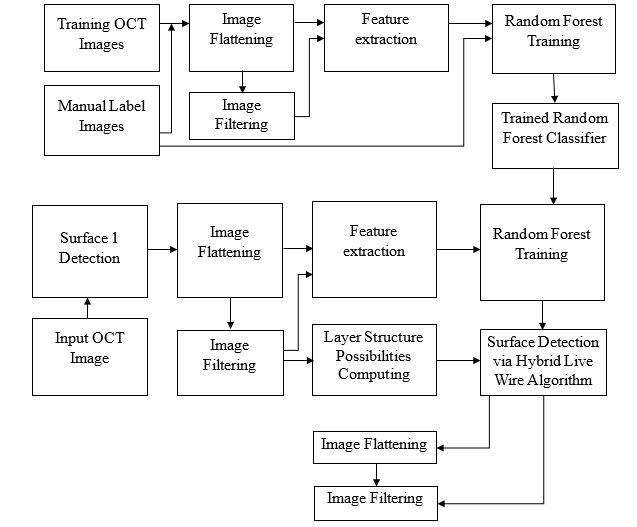
The proposed framework can be split into two stages: training stage and testing stage. In the training stage, OCT images are manually annotated and features are extracted for random forest classifier training. In the testing stage, the proposed segmentation method is a coarse-to-fine segmentation process that consists of three steps: preprocessing, initialization and segmentation. Original OCT image is preprocessed to reduce noise and gray levels are normalized. The necessary feature vector is computed from preprocessed OCT images and initial surfaces of retinal layer are computed with the application of random forest classifiers to label voxels as different retinal layers. The final surfaces are refined via the proposed hybrid live wire algorithm and fluid is also segmented.
BLOCK DIAGRAM