- ALL COMPUTER, ELECTRONICS AND MECHANICAL COURSES AVAILABLE…. PROJECT GUIDANCE SINCE 2004. FOR FURTHER DETAILS CALL 9443117328
Projects > ELECTRONICS > 2019 > IEEE > DIGITAL IMAGE PROCESSING
Melanoma is considered a fatal type of skin cancer. However, it is sometimes hard to distinguish it from Nevus due to their identical visual appearance and symptoms. The mortality rate because of this disease is higher than all other skin related consolidated malignancies. The number of cases is growing amongst young people but if it is diagnosed at its earlier stage then the survival rates become very high. The cost and time required for the doctors to diagnose all patients for Melanoma are very high. In this research work, we propose an intelligent system to detect and distinguish Melanoma from Nevus by using state of the art image processing techniques. At first, Gaussian Filter is used for removing noise from the skin lesion of the acquired images followed by the use of improved K-mean clustering to segment out the lesion. A distinctive hybrid super feature vector is formed by the extraction of textural and color features from the lesion. Support Vector Machine (SVM) is utilized for the classification of skin cancer into melanoma and nevus. Our aim is to test the effectiveness of the proposed segmentation technique, extract the most suitable features and compare the classification results with the other techniques present in the literature. The proposed methodology is tested on DERMIS dataset having a total number of 397 skin cancer images where 146 are melanoma and 251 are nevus skin lesions. Our proposed methodology archives encouraging results having 96% accuracy.
Edge based detection method, filtering methods
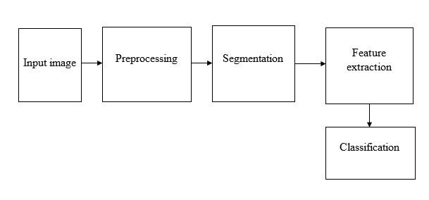
Input images acquired from the datasets undergo quality enhancement through preprocessing techniques. Later the ROI from the skin lesion is extracted which are further processed for significant feature extraction and lastly classifying them into melanoma or nevus.  
BLOCK DIAGRAM