- ALL COMPUTER, ELECTRONICS AND MECHANICAL COURSES AVAILABLE…. PROJECT GUIDANCE SINCE 2004. FOR FURTHER DETAILS CALL 9443117328
Projects > ELECTRONICS > 2019 > IEEE > DIGITAL IMAGE PROCESSING
A tumor cell is a form of cell that develops out of control of the ordinary forces and standardizes growth. Brain tumor is one of the major reasons for human death every year. Around 50% of brain tumor diagnosed patient die with primary brain tumors each year in the United States. Electronic modalities are used to diagnose brain tumors. Among all electronic modalities, Magnetic Resonance Imaging (MRI) is one of the most used and popular for brain tumor diagnosis. In this research study, an automated approach has been proposed where MRI gray-scale images were incorporated for brain tumor detection. This study proposed an automated approach that includes enhancement at the initial stage to minimize gray-scale color variations. Filter operation was used to remove unwanted noises as much as possible to assist better segmentation. As this study test grayscale images therefore; threshold based OTSU segmentation was used instead of color segmentation. Finally, pathology experts provided feature information was used to identify the region of interests (brain tumor region). The experimental results showed that the proposed approach was able to perform better results compared to existing available approaches in terms of accuracy while maintaining the pathology experts’ acceptable accuracy rate.
Sobel, Prewitt and Canny edge detection, k-means clustering
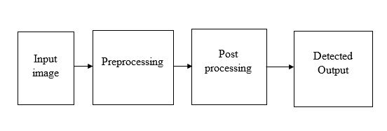
MRI image acquisition is the first step of the method. The identification of brain tumor using MRI images includes two processes: pre-processing and post-processing. A number of steps incorporated in this stage which include: a. Image Enhancement, b. Filter Operation, c. Segmentation. Image enhancement is used as the first step of image preprocessing for this study. This study used contrast stretching as it comparatively performs better on the gray scale image as contrast increased without distorting relative gray level intensities. Filter operation is performed on the image to increase the smoothness, sharpness as well as edge enhancement. Median filter has been applied in the proposed method. Segmentation divides the image into regions based on the similar attributes. Thresholding based Otsu’s method is used of its wide uses to segment an image for further processing like feature analysis as well as to do the binary transformation of an image. Post-processing includes: a. Feature Extraction, b. Identification. This system uses four properties which include area, circularity (roundness and diameter), and solidity are act perform the work of feature extraction and also applied PCA feature selection algorithm to verify that the selected features are appropriate to detect the ROI (region of interest). The aim of identification process is to detect the ROI. The ROI is detected by using the features extracted in the feature extraction section.
BLOCK DIAGRAM