- ALL COMPUTER, ELECTRONICS AND MECHANICAL COURSES AVAILABLE…. PROJECT GUIDANCE SINCE 2004. FOR FURTHER DETAILS CALL 9443117328
Projects > ELECTRONICS > 2019 > IEEE > DIGITAL IMAGE PROCESSING
In the present scenario retinal image processing is toiling hard to get an efficient algorithm for de-noising and segmenting the blood vessel confined inside the closed curvature boundary. On this ground, this paper presents a hybrid active contour model with a novel preprocessing technique to segment the retinal blood vessel in different fundus images. Contour driven black top-hat transformation and phase based binarization method has been implemented to preserve the edge and corner details of the vessels. In the proposed work, Gradient Vector Flow (GVF) based snake and balloon method are combined to achieve better accuracy over different existing active contour models. In the earlier active contour models, the snake cannot enter inside the closed curvature resulting loss of tiny blood vessels. To circumvent this problem, an inflation term ð…ð¢ð§ðŸ(ð›ðšð¥ð¥ð¨ð¨ð§) with GVF based snake is incorporated together to achieve the new internal energy of snake for effective vessel segmentation. The evaluation parameters are calculated over four publically available databases: STARE, DRIVE, CHASE and VAMPIRE. The proposed model outperforms its competitors by calculating a wide range of proven parameters to prove its robustness.
Canny edge detection and KNN
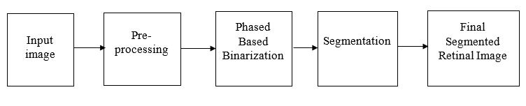
In this method of proposed segmentation, a pre-processed image is considered as the input to the hybrid active contour algorithm. In the outset, an inverted green channel image is extracted from RGB channelled fundus image. the image obtained from the inverted green channel is enhanced in the first step only. This approach has enhanced the minute blood vessels but at the same time noise present in the image was also enhanced. This noise was further removed in main binarization process. For the enhancement, contrast limit adaptive histogram equalization (CLAHE) is applied. As a result, a small flash of light appears at the center of the vessel, known as central vessel reflex. To eliminate this, a contour driven black top-hat transform algorithm is applied on vessel enhanced image. This is the last algorithm applied over the image to get final pre-processed image. Further, the resultant image from this step is segmented by hybrid active contour segmentation algorithm. Filtering of noise, obtained so far, is done using Kovesi phase preserved denoising method. This approach has hybridized the conventional snake model resulting in better segmentation of blood vessels.