- ALL COMPUTER, ELECTRONICS AND MECHANICAL COURSES AVAILABLE…. PROJECT GUIDANCE SINCE 2004. FOR FURTHER DETAILS CALL 9443117328
Projects > ELECTRONICS > 2020 > IEEE > DIGITAL IMAGE PROCESSING
We introduce an approach for image segmentation based on sparse correspondences between keypoints in testing and training images. Keypoints represent automatically identified distinctive image locations, where each keypoint correspondence suggests a transformation between images. We use these correspondences to transfer label maps of entire organs from the training images to the test image. The keypoint transfer algorithm includes three steps: (i) keypoint matching, (ii) voting-based keypoint labeling, and (iii) keypoint-based probabilistic transfer of organ segmentations. We report segmentation results for abdominal organs in whole-body CT and MRI, as well as in contrast-enhanced CT and MRI. Our method offers a speed-up of about three orders of magnitude in comparison to common multi-atlas segmentation, while achieving an accuracy that compares favorably. Moreover, keypoint transfer does not require the registration to an atlas or a training phase. Finally, the method allows for the segmentation of scans with highly variable field-of-view.
Scale invariant feature transform (SIFT)
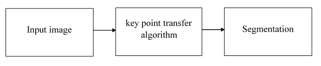
The proposed system introduces an approach for image segmentation based on sparse correspondences by identifying distinctive locations in the image: keypoints. Keypoints are automatically computed as local optima of a saliency function, contrary to manually selected landmarks. We match keypoints between test and training images to establish correspondences for a sparse set of image locations. Based on these correspondences, the segmentation masks of entire organs are transferred and fed into a probabilistic fusion algorithm. The segmentation accuracy compares favorably to common multi-atlas techniques, while working with sparse correspondences leads to a computationally efficient algorithm, offering orders of magnitude of speed-up. We outline the keypoint transfer segmentation algorithm and as animation in the supplementary material. Keypoints are extracted at salient image regions and described by their geometry and a descriptor based on a histogram of local image intensity gradients. Following keypoint extraction, we segment an image in three steps. First, we match keypoints in the test image to keypoints in the training images based on the geometry and the descriptor. Second, keypoint labels are voted on based on matches. The keypoint receives two votes for right kidney and one for liver, resulting in a majority vote for right kidney. Third, the label mask is transferred for the entire organ for each match that is consistent with the majority label vote. The organ map from one training image is possibly transferred multiple times if more than one match is available for this training image. Keypoint transfer also integrates the certainty in the keypoint label voting and computes the intensity similarity between scans. The algorithm’s capability in approximating the organ shape can further improve with a growing number of manually labeled scans, where additional images can be included in the training set without the need for a dedicated training stage.
BLOCK DIAGRAM