- ALL COMPUTER, ELECTRONICS AND MECHANICAL COURSES AVAILABLE…. PROJECT GUIDANCE SINCE 2004. FOR FURTHER DETAILS CALL 9443117328
Projects > ELECTRONICS > 2020 > IEEE > DIGITAL IMAGE PROCESSING
Automated and accurate segmentation of cystoid structures in Optical Coherence Tomography (OCT) is of interest in the early detection of retinal diseases. It is however a challenging task. We propose a novel method for localizing cysts in 3D OCT volumes. The proposed work is biologically inspired and based on selective enhancement of the cysts, by inducing motion to a given OCT slice. A Convolutional Neural Network (CNN) is designed to learn a mapping function that combines the result of multiple such motions to produce a probability map for cyst locations in a given slice. The final segmentation of cysts is obtained via simple clustering of the detected cyst locations. The proposed method is evaluated on two public datasets and one private dataset. The public datasets include the one released for the OPTIMA Cyst segmentation challenge (OCSC) in MICCAI 2015 and the DME dataset. After training on the OCSC train set, the method achieves a mean Dice Coefficient (DC) of 0.71 on the OCSC test set. The robustness of the algorithm was examined by cross validation on the DME and AEI (private) datasets and a mean DC values obtained were 0.69 and 0.79, respectively. Overall, the proposed system has the highest performance on all the benchmarks. These results underscore the strengths of the proposed method in handling variations in both data acquisition protocols and scanners.
Topographical Watershed Algorithm
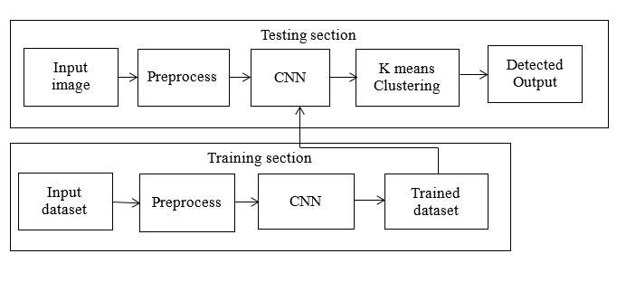
The proposed pipeline consists of three main stages, namely, pre-processing, detection, and segmentation via clustering. SD-OCT volumes are generally acquired with different operator-defined protocols. The signal from the SD-OCT volumes are usually affected by speckle noise. Speckle noise is signal dependent and hence depends on the structure of the tissue in an OCT volume. A graph-based segmentation approach is used to extract only the ILM and RPE layers to form a mask. Selective enhancement is done by employing the notion of Generalized Motion Pattern (GMP). Next, we construct an ensemble of these GMPs and learn a function (using a CNN) to combine them such that only cysts are enhanced. The trained CNN model learns a function ? that combines the cake of GMP C to produce a probability map with cyst pixels having higher probability score than background pixels. Due to the smearing effect of GMP, a rough region around the objects of interests (cysts), rather than accurate cyst regions, are detected. Thus, segmentation is required to obtain a precise cyst boundary. The probability map is thresholded to obtain a binary map representing detected cyst regions. This map is multiplied with the ROI image to extract the detected regions in the intensity space. K-means clustering is applied on this product image. Detected regions are clustered into cysts, false positives region and background in the intensity space. Since cysts are relatively darker regions compared to the false positive regions, the clusters with lower mean intensity are retained as desired cyst segments.
BLOCK DIAGRAM